News
Artificial intelligence analyzes whole mouse bodies down to cell level
20 May 2026
A team led by LMU researcher Ali Ertürk finds evidence of previously unknown nerve damage in obesity.

20 May 2026
A team led by LMU researcher Ali Ertürk finds evidence of previously unknown nerve damage in obesity.
An international research team led by LMU Professor Ali Ertürk* has developed a new AI-powered method that enables whole-body analysis of mice down to the single-cell level. Helmholtz Munich and LMU served as the lead institutions for the project, which also involved additional researchers worldwide. Dubbed “MouseMapper,” the novel system combines advanced tissue clearing, light-sheet microscopy, and deep learning to automatically map neural pathways, immune cells, and 31 different organs and tissues throughout the body, as the researchers report in the journal Nature.
“For the first time, MouseMapper allows us to visualize pathological changes at the cellular level throughout the entire body,” says Professor Ali Ertürk from the Institute for Stroke and Dementia Research at LMU and Helmholtz Munich. “This opens up entirely new possibilities for detecting diseases at very early stages – long before the first symptoms appear.”
Ertürks team is making the complete whole-body data online available to other researchers worldwide.
„For the first time, MouseMapper allows us to visualize pathological changes at the cellular level throughout the entire body. This opens up entirely new possibilities for detecting diseases at very early stages – long before the first symptoms appear.“Professor Ali Ertürk, Institute for Stroke and Dementia Research at LMU
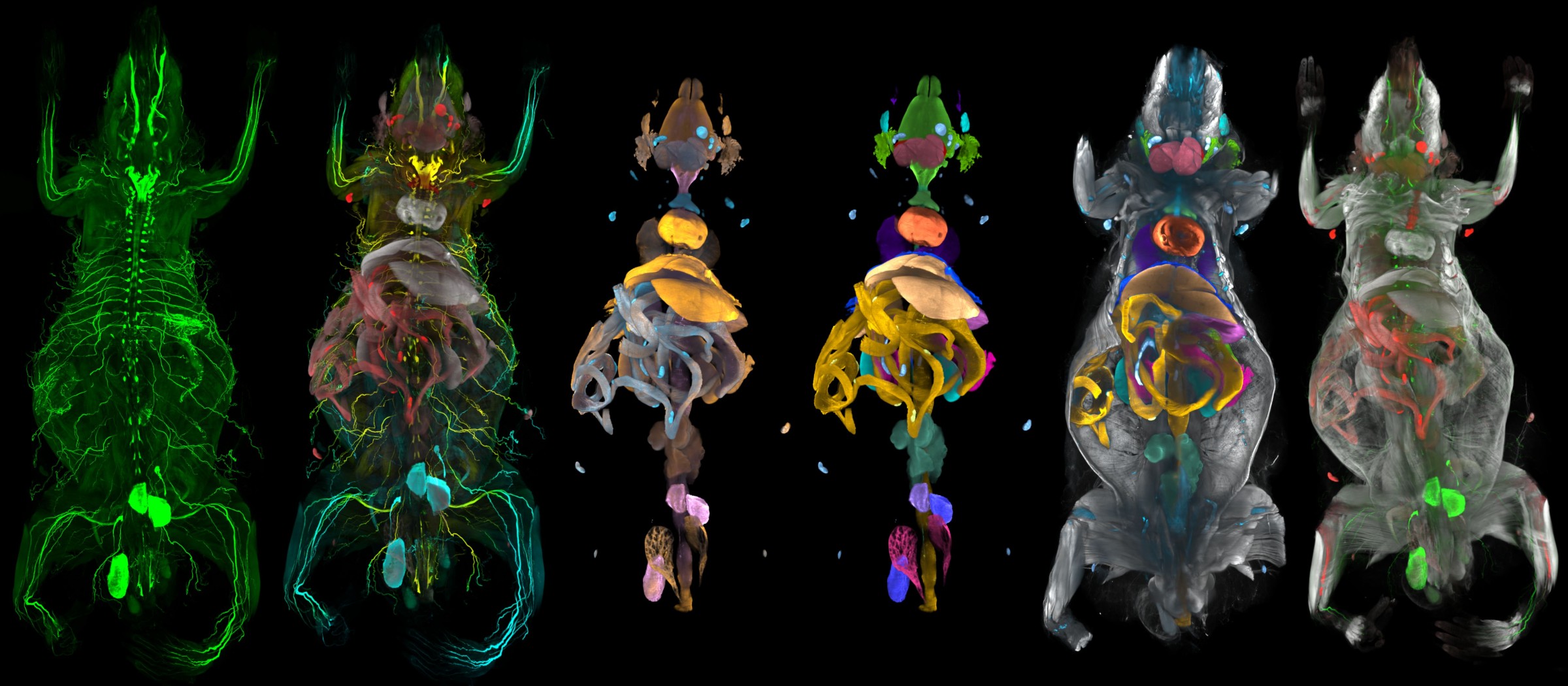
MouseMapper automatically segments 31 organs and tissue types in a mouse while simultaneously mapping neural and immune cells throughout the body. This enables comprehensive multi-organ analyses in intact mice.
© Ertürk Lab | Helmholtz MunichTechnologically, MouseMapper is based on so-called foundation models – large AI models that are trained on vast datasets to recognize general patterns. Unlike classical AI systems, they are not built for a single task, but can be flexibly adapted to many different applications. Accordingly, the system does not just work for a certain disease or imaging method, but can be flexibly applied to other datasets.
The AI automatically recognizes the finest nerve structures, accumulations of immune cells, and anatomical regions throughout the body. Researchers can thus systematically compare changes and pick out conspicuous regions for further molecular analyses.

Prof. Ali Ertürk | © Helmholtz Munich | Matthias Tunger
The researchers used MouseMapper, among other things, to investigate the effects of obesity on the entire organism of mice. In the process, they discovered previously unknown structural damage to sensitive facial nerves of the trigeminal system. This particularly affected the infraorbital nerve, which is responsible for tactile perception via the whiskers. Mice with fatty diets had significantly fewer nerve branches and responded worse to neural stimuli.
Additional molecular analyses revealed changes in signaling pathways associated with inflammations and remodeling and degeneration processes of nerve cells. It was particularly noteworthy that scientists had already found the same molecular patterns in tissue samples from humans with obesity. This established the first direct connection between observations in the mouse model and changes in humans.
“Our finding that obesity can clearly damage sensory facial nerves was particularly surprising,” explains Dr. Doris Kaltenecker, 1st author of the study. “The fact that the same molecular patterns were found in humans makes the results all the more relevant.”
In addition to nerve damage, MouseMapper documented extensive inflammatory processes in various organs of obese animals. Fatty tissue, the liver, and the abdominal cavity were most strongly affected. The researchers found larger accumulations of immune cells in these areas – an indication of chronic inflammatory responses throughout the organism.
The long-term goal of Ertürk’s team is to create comprehensive cell atlases of healthy and diseased organisms. “We want to build digital cell atlases of the body that function like virtual twins,” says the scientist. “In the future, this could enable us to analyze diseases on computers and test novel therapies much faster than is currently the case.”
Digital twins are virtual models of biological organisms that simulate the body as realistically as possible at the cellular level using AI, imaging, and molecular data. This could allow researchers to simulate diseases, digitally test therapies, and better understand pathological processes in the future – even without additional animal experiments in some cases.
Doris Kaltenecker, Izabella Horvath, Rami Al-Maskari, Ali Ertürk et. al. A deep-learning framework reveals whole-body perturbations at cell level. Nature. 2026