Forgetting visualized in brain cells
28 Oct 2022
LMU physicist Paola Coan presents new imaging technique that allows brain cells of mice suffering from Alzheimer's disease to be visualized in previously unattained quality.
28 Oct 2022
LMU physicist Paola Coan presents new imaging technique that allows brain cells of mice suffering from Alzheimer's disease to be visualized in previously unattained quality.

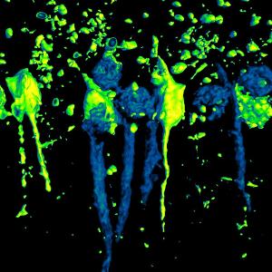
3D rendering of phase-contrast X-ray tomography of Alzheimer neurons (green) and healthy cells (blue). | © Paola Coan / LMU
There is a growing understanding of what happens in the brain when it is afflicted with Alzheimer's disease. Due to the death of nerve cells, people with Alzheimer's become increasingly forgetful, confused and disoriented. To date, Alzheimer's is incurable. But there are many approaches to getting the disease under control. One of these is an understanding of the neuronal processes that take place in our thinking organ. A team led by Prof. Paola Coan from the Department of Medical Physics and the Department of Radiology at the LMU Hospital in Munich has now gained new findings concerning this process with the help of X-ray phase-contrast computed tomography. Using this imaging technique, the scientists have gained unique insights into the ageing and neurodegeneration of brain cells in mice suffering from Alzheimer's disease.
Giacomo E. Barbone, Alberto Bravin, Alberto Mittone, Alexandra Pacureanu, Giada Mascio, Paola Di Pietro, Markus J. Kraiger, Marina Eckermann, Mariele Romano, Martin Hrabě de Angelis, Peter Cloetens, Valeria Bruno, Giuseppe Battaglia & Paola Coan. X-ray multiscale 3D neuroimaging to quantify cellular aging and neurodegeneration postmortem in a model of Alzheimer’s disease. European Journal of Nuclear Medicine and Molecular Imaging, 2022.