“Friends and Foes”: exhibition by PlantMicrobe research alliance
30 Oct 2023
Microphotographs show how microbes and plants interact – for good and ill.
30 Oct 2023
Microphotographs show how microbes and plants interact – for good and ill.

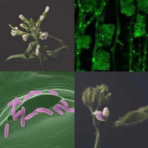
The relationship between microorganisms and plants is multifaceted. | © LMU, Jessica Folgmann, MPI für Biologie. Collage: LMU
From partners to mortal enemies – the relationship between microorganisms and plants is multifaceted. The exhibition “Friends and Foes” at Munich Botanical Garden, which opens on 4 November, illustrates these manifold interactions in photographs created by researchers from the Collaborative Research Centre TRR 356 “PlantMicrobe”.
Millions of bacteria and fungi live on and in each and every plant. Some improve the supply of nutrients or protect against pests. By contrast, pathogenic bacteria or fungi can cause massive damage to the plant, which tries to protect itself by means of natural defenses and a sophisticated immune system. Whether useful or harmful, interactions affect plant health and thus impact crop yields. The Collaborative Research Centre “PlantMicrobe” investigates the molecular mechanisms that determine plant-microbe symbioses and the genetic diversity of the actors. “The better we understand these interactions and the interplay between them, the more scope we will have to ensure humankind has an adequate supply of healthy food,” says Dr. Dagmar Hann, scientist at LMU’s Biocenter and co-organizer of the exhibition. The spokesperson of the research center is Professor Martin Parniske, Head of Genetics at the Biocenter.
In 19 photographs, students and scientists in TRR 356 offer a microscopic glimpse into biological processes that are not visible to the naked eye. The images depict phenomena such as cellular changes wrought by the symbiosis of plants and bacteria and the infection strategies of microorganisms. These processes are magnified using optical and laser microscopes and made visible with the aid of dyes.